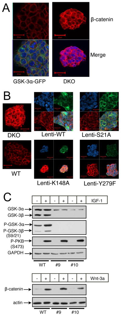
Figure 4.

Rescue of DKO ES cells with variants of GSK-3α. (A) Immunofluorescent images of DKO ES cells and DKO ES cells reconstituted with a GSK-3α-GFP fusion protein. β-catenin staining is red, GFP staining is green and nuclear staining is blue (DAPI). (B) Immunofluorescent images of WT and DKO ES cells as well as DKO ES cells rescued with various V5-epitope-tagged mutants of GSK-3α through lentiviral transduction. V5 staining is green, β-catenin staining is red and nuclear staining is blue. All images were obtained with identical PMT, resolution and scan-rate settings. (C) DKO ES cells rescued with a GSK-3α S21A mutant still respond to Wnt-3a. #9 and #10 are independent clones of DKO ES cells reconstituted with S21A-GSK-3α. Treatments with IGF-1 were for 15 minutes, while treatments with Wnt-3a conditioned medium were overnight.
