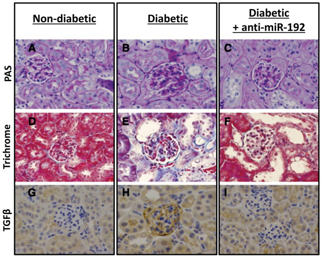
Figure 5.
LNA–anti-miR-192 attenuates glomerular growth, mesangial expansion, and TGFβ expression in 17-week diabetic mice. (A-C) Periodic acid–Schiff [PAS] staining of representative kidney sections. (D-F) Masson’s trichrome staining showing glomerular and tubulointerstitial fibrosis. (G-I) TGF-β immunostaining of kidney sections. Reproduced from Putta et al. (2012); with kind permission of the Journal of the American Society of Nephrology.

