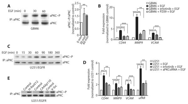
Fig. 2. aPKC is activated downstream of tumor-intrinsic oncogenic RTK signaling pathways.
(A) Activation of aPKC in GBM6 cells after EGF treatment for the indicated times. Active aPKC was detected after aPKC immunoprecipitation followed by aPKC phosphoThr410/403 immunoblotting. The blot was stripped and reprobed with aPKC. Representative (left) and quantified data from all experiments (right) are presented. (B) Expression of the indicated genes in GBM6 cells upon treatment with EGF alone or in the presence of PZ09 or erlotinib, as detected by RT-qPCR and normalized to GBM6 control cells. (C) Activation of aPKC in U251/EGFR cells after EGF treatment for the indicated times. (D) Expression of the indicated genes in U251/EGFR cells, U251/EGFR cells treated with erlotinib, or U251/EGFR cells transfected with aPKC siRNA and treated with EGF, as detected by RT-qPCR and normalized to U251/EGFR control cells. (E) Activation of aPKC in U251/EGFR cells after transfection with vector alone or active Ras (H-RasV12), active PI3K (CAAXp110), or active Src (v-Src). Data for (A) to (E) are presented as representative images or as means ± SEM of at least three independent experiments. *P < 0.05, **P < 0.01, ***P < 0.001.

