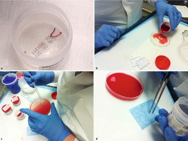
Fig. 2.

Tissue was expressed into the formalin container. a Visible core. b More bloody specimens; the contents of the container were poured into a petri dish. c The formalin-fixed pieces of liver tissue, distinguished from blood clot, were removed with forceps. d Next, the tissue was wrapped in lens paper and placed into a histology cassette for standard processing; the blood clot was also submitted. Usually, all material was processed in a single cassette.
