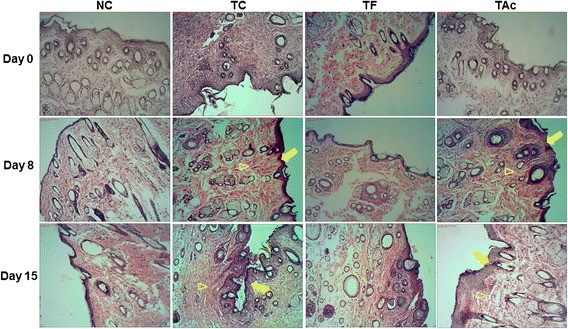
Fig. 1.

The histological evaluation of the skin flaps revealed by HE coloration (10x). Microscopic examination of TC and TAc groups indicated regression of the lesions with better epithelialization (arrows) and more effective re-organization of the dermis (arrowhead) compared to the NC and TF on 8th and 15th days after injury
