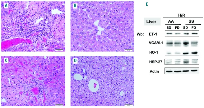
Figure 5.

FD reduces H/R-induced liver damage and vascular activation. (A–D) Hematoxylin and eosin-stained sections of liver tissue at 400x magnification from SS mice under a SD (A and C) or FD supplementation (B and D) exposed to H/R. Livers from mice given FD have less inflammatory cellular infiltrate (B) and thrombi (D) than livers from SS mice fed a SD. The infiltrate shown best in (B) is composed mostly of lymphocytes. Scattered hemosiderin deposits and areas of necrosis are also present. (E) Immunoblot analysis with specific antibodies against ET-1, VCAM-1, HO-1, and heat shock protein-27 (HSP27) of liver from AA and SS mice treated as in (A–D). One representative gel from six with similar results is presented. Densitometric analysis of ET-1, VCAM-1, HO-1 and HSP27 immunoblots is shown in Online Supplementary Figure S5.
