
Figure 3.
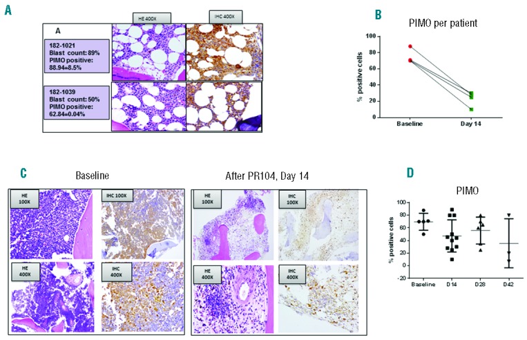
(A). Examples of hematoxylin and eosin (HE) staining (left) and PIMO immunohistochemical (IHC) staining (right) in bone marrow (BM) biopsy specimens from 2 representative patients. Original magnification, ×400. (B). Quantification of PIMO-positive cells in BM before and after PR104 (day 14) treatment by CRi spectral imaging and Inform software analysis (=4). None of these patients achieved an objective response. (C). Example of paired BM biopsy specimens (before and after PR104) in patient 182-1017 (ALL). Baseline BM blasts, 98%; day 14, 43%. Fraction of PIMO-positive cells: baseline, 82%; day 14, 28%. Original magnification is shown in the boxed areas. (D). Fraction of PIMO-positive cells in all BM specimens tested.