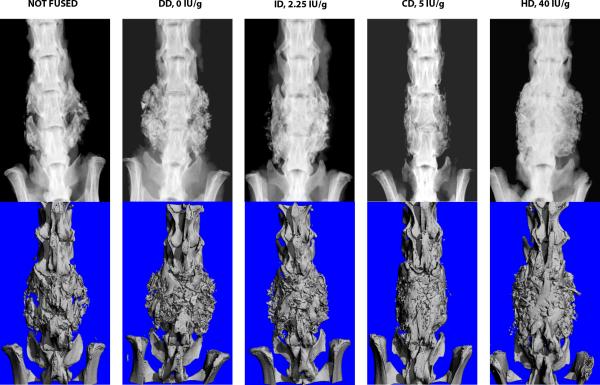
Figure 5.
Example high resolution radiographs (top) with μCT (below) of ex vivo specimens from each of the vitamin D-adjusted chow groups. Robust radiographic fusion is observed in the High Vitamin D (40 IU/g) specimen. A radiographic example of ‘not fused’ is included for comparison (left most radiograph).

