
FIGURE 8.
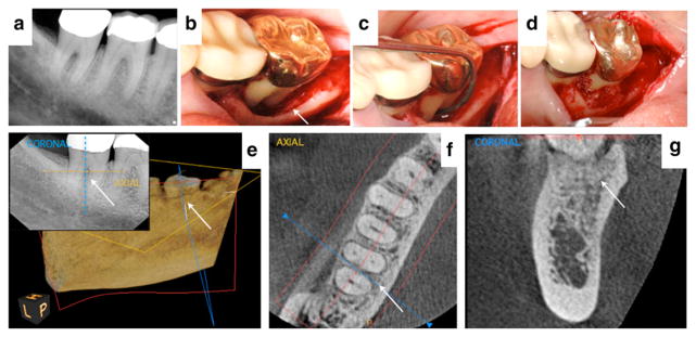
A healthy 55-year-old female with 9-mm probing depths and Class II furcation involvement on the lingual aspect of tooth #31 (case courtesy of BSM). 8a Preoperative radiograph showing bone loss in the furcation. 8b and 8c The extensive size of the periodontal defect can be visualized after flap reflection. 8d The cellular allograft|||| containing mesenchymal stem cells and osteoprogenitor cells was delivered into the periodontal defect. 8e A 6-month two-dimensional radiograph and three-dimensional cone-beam computed tomorgraphy¶¶ (CBCT) view of the site after 3 years suggests significant bone fill. Color dotted lines on the cross-sectional radiograph assist location of the coronal and axial views from the CBCT. 8f and 8g Axial and coronal slices from a 3-year postoperative CBCT reflect complete bone fill of the furcation and supportive evidence of a normal periodontal ligament space (5 × 5 cm scan set at 90-μm resolution). Figures 8a, 8c, and 8d reproduced with permission from Quintessence (McAllister6).