
Figure 1. Orthotopic AT-3-FL tumor implants in CXCR7ΔEND/ΔEND mice have more viable tumor cells.
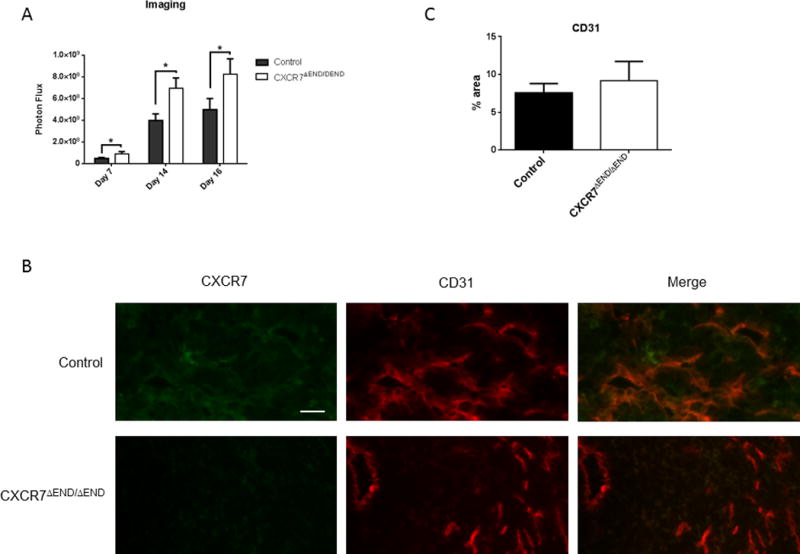
A) Growth of AT-3-FL breast cancer cells implanted orthotopically into CXCR7ΔEND/ΔEND and control mice monitored by bioluminescence imaging at indicated days (n = 8 mice per group). Graph depicts mean values + SEM for photon flux. *, p < 0.05. B) Immunofluorescence of excised orthotopic AT-3-FL tumors for CD31-tumor vasculature (green) and CXCR7 (red). White arrows show co-localization of CD31 and CXCR7 in merged images. AT-3-FL cells express CXCR7, accounting for staining throughout both tumors. Scale bar designates 50 μm. C. Graph shows mean values + SEM for area occupied by CD31+ blood vessels in tumors from each group of mice.