
Figure 5. AT-3-FL cells produce greater experimental metastases in CXCR7ΔEND/ΔEND mice.
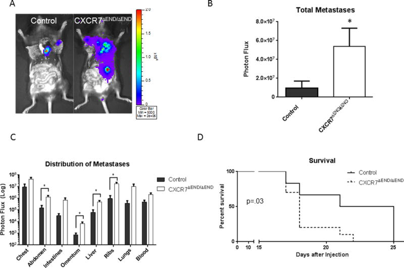
A) We injected AT-3-FL cells systemically via injection into left ventricles of CXCR7ΔEND/ΔEND and control mice. Presented images are representative of relative amounts of metastatic AT-3-FL cells at the time of euthanization. B) We quantified total AT-3-FL metastases by bioluminescence imaging and graphed data as mean values + SEM for each group (n = 12 each). C) We determined site-specific localization of metastases by bioluminescence imaging and quantified tumor burden as mean values + SEM for photon flux in each site. D) Survival curves for a separate experiment in which AT-3-FL cells were injected into CXCR7ΔEND/ΔEND and control mice and monitored until animals were euthanized for humane endpoints (n = 12 per group). * p < 0.05.