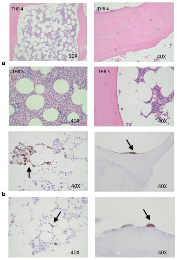
Figure 3.

Breast cancer cell colonization of bone tissue. (a) H & E stained histological sections of human bone tissue specimens before culture. (b) Immunohistochemical staining of bone fragments after co-culture with MDA-MB-231-fLuc cells for 72 h with cytokeratin antibody reveals breast cancer cell colonization of the marrow compartment (left panels), and attachment to ossified bone surfaces (right panel).
