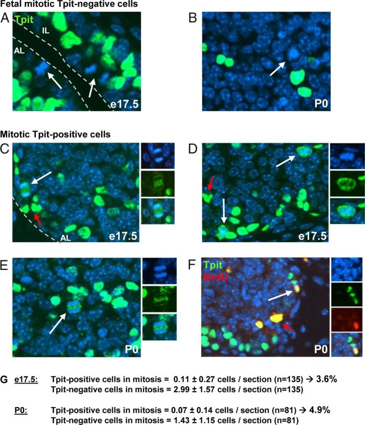
Figure 1.

Division of Differentiated Corticotrope Cells. A–E, e17.5 and P0 mouse pituitary sections were subjected to immunofluorescence using Tpit antibody (green) and nuclei counterstained with Hoechst (blue). A and B, Most dividing cells (arrows) are Tpit negative and located around the pituitary lumen (A). Tpit-negative mitotic cells are also observed within the AL (B). C–E, Mitotic Tpit-positive cells (white arrow) are also observed, and Tpit immunoreactivity (green) is excluded from condensed chromatin (blue). Red arrows indicate background autofluorescence by red blood cells. F, Colabeling of P0 pituitary sections for Tpit (green) and BdrU (blue). Tpit and BrdU colabeling indicates recent passage through S phase. G, Count of mitotic Tpit-positive and negative cells in e17.5 and P0 pituitary sections.
