Figure 5.
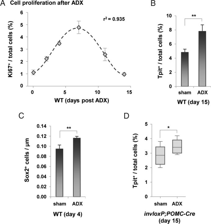
Assessment of Progenitor and Corticotrope Expansion after ADX. A, Progenitor cell proliferation in pituitary following ADX was assessed using Ki67 immunostaining and found to follow a time course that is similar to that observed by Nolan et al. (21). The number of proliferating cells peaks at day 7 and is back to baseline after 14 days. B, Increase of Tpit-positive cells in ADX mice 15 days after surgery compared with sham-operated controls. C, Quantification of Sox2-positive cells along the pituitary cleft 4 days after surgery in sham and ADX mice (10 wk old). Note that the abundance of Sox2-positive cells is higher in these mice compared with the 37-week-old mice of Figure 4D: this reflects an age-dependent depletion of Sox2-positive cells. D, Proportion (%) of Tpit-positive corticotropes in AL 15 days after ADX compared with sham-operated mice. Despite a 40% corticotrope cell loss in the 26-week-old invloxP/+;POMC-Cre mice used for these experiments, the significant increase of corticotropes is consistent with expansion of progenitors followed by differentiation rather than self-duplication of corticotropes. Data are presented as means ± SEM (n = 3–7). * and ** indicate P ≤ .05 and P ≤ .01, respectively.

