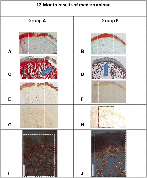
Fig. 5.

Histological evaluation of osteochondral defects 12 months postop (original magnification of (a–h) ×12, white dashed box is the osteochondral defect outline). a A proteoglycan-specific dye stains the cartilage layer in a group A animal. The osteochondral defect is still not reconstructed with eburnated bone at the joint surface. Note cartilage disruption, cleft formation, cartilage matrix loss. disrupted subchondral bone plate, and irregular bone trabeculae (Safranin-O stain, Fast Green counterstain). b A group B animal showing normal cartilage. c A bone cyst is seen in a group A animal (arrowhead), with massive thickening of the subchondral bone plate (Masson Trichrome stain). d A group B animal demonstrates perfect repair of the subchondral bone underlying the regenerated cartilage (arrowhead) (Masson Trichrome stain). e Collagen type II staining reveals lack of collagen type II in some of the arthritic tissue overlying the osteochondral defect with some staining in the bone cyst indicating heterotopic cartilage formation in a group A animal (anti-collagen type I antibody). f Collagen type II stain is uniform in the newly created cartilage in a group B animal; there is no collagen type II staining in the regenerated subchondral bone (anti-collagen type II antibody). g Collagen type I stain is uniform in the bone, but there are areas within the articular cartilage that stain positively as well in a group A animal (anti-collagen type I antibody). h Uniform collagen type I stain in a group B animal (anti-collagen type I antibody). i Disordered repair collagen fibrils are evident in this polarized light microscopy image of a group A animal and lack of repair tissue zonation (original magnification of this and j was ×40). (j) Well-ordered mature collagen fibrils are evident within the subchondral bone in a group B animal. The cartilage layer demonstrates a clear zonation phenomenon
