
Fig. 1.
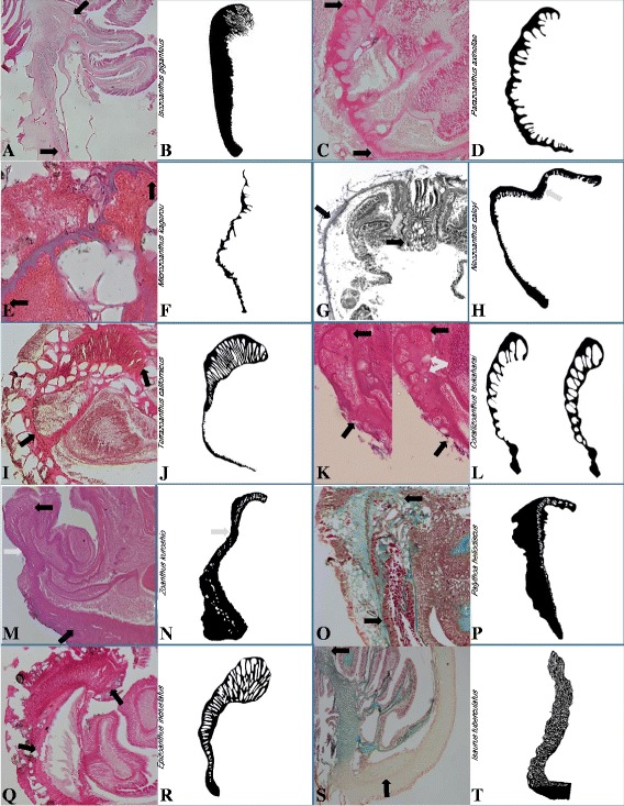
Marginal musculature arrangements. Photographs of histological section of marginal musculature (between black arrows) with accompanying drawing of isolated mesogleal structures supporting the marginal musculature for branchiform endodermal (Isozoanthus giganteus: a, b), cteniform endodermal (Parazoanthus axinellae: c, d), spindly-cteniform endodermal (Microzoanthus kagerou: e, f), discontiguous endodermal (Neozoanthus caleyi: g, h; grey arrow indicates undifferentiated mesoglea; histological image reproduced with permission of J. Reimer), meso-endo transitional (Terrazoanthus californicus: i, j), cyclically transitional (Corallizoanthus tsukaharai: k, l; grey arrows indicate lacunae formed by dissolution of foraminifera), discontiguous mesogleal (Zoanthus kuroshio: m, n; grey arrow indicates undifferentiated mesoglea), linear mesogleal (Palythoa heliodiscus: o, p), reticulate mesogleal (Epizoanthus incrustatus: q, r), orthogonally-reticulate mesogleal (Isaurus tuberculatus: s, t)