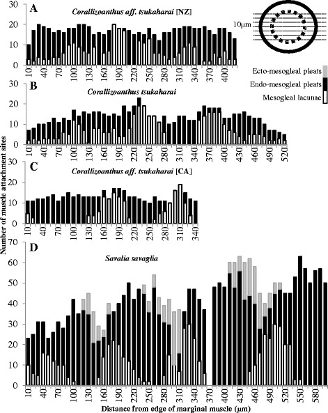
Fig. 2.

Patterns in the muscle attachment sites of cyclically transitional marginal muscle arrangements. Number, position, and type of marginal muscle attachment sites as they appear within serial longitudinal sections of Corallizoanthus aff. tsukaharai [NZ] (a), Corallizoanthus tsukaharai (b), Corallizoanthus aff. tsukaharai [CA] (c), and Savalia savaglia (d). Each bar represents a 10 μm longitudinal section with the number and type of muscle attachment points; gray bars indicate ectoderm-facing mesogleal pleats, black bars indicate endoderm-facing mesogleal pleats, open bars indicate mesogleal lacunae. Empty positions indicate data missing due to sectioning artifact. Inlay diagram demonstrates plane of microtome blade (dotted lines) against the diameter of the polyp (outer ring) and marginal muscle (broken ring)
