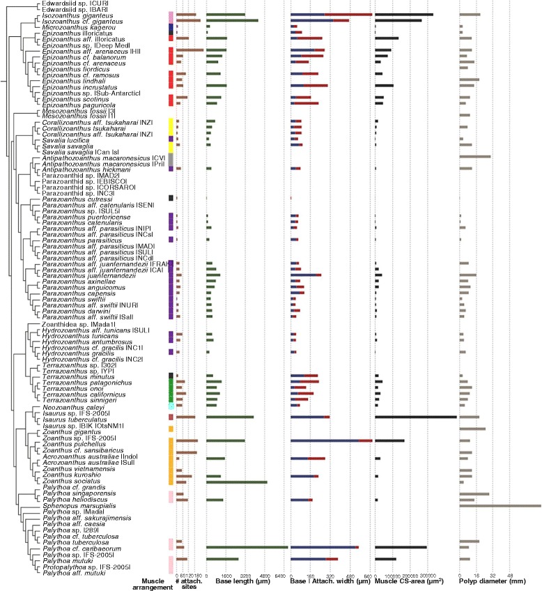
Fig. 8.

Marginal musculature arrangements and the dimensions of their structural components mapped to the composite phylogeny. Boxes indicate muscle arrangement character state (branchiform endodermal [purple], cteniform endodermal [violet], spindly-cteniform endodermal [blue], discontiguous endodermal [light blue], meso-endo transitional [green], cyclically transitional [yellow], discontiguous mesogleal [orange], linear mesogleal [pink], reticulate mesogleal [red], orthogonally-reticulate mesogleal [burgundy], endo-meso transitional [grey], simplified mesogleal [black]); bar graphs indicate the mean maximum number of attachment sites, mesogleal base length (μm), mesogleal base and attachment site width (μm), marginal muscle cross-sectional area (μm2), and polyp diameter (mm)
