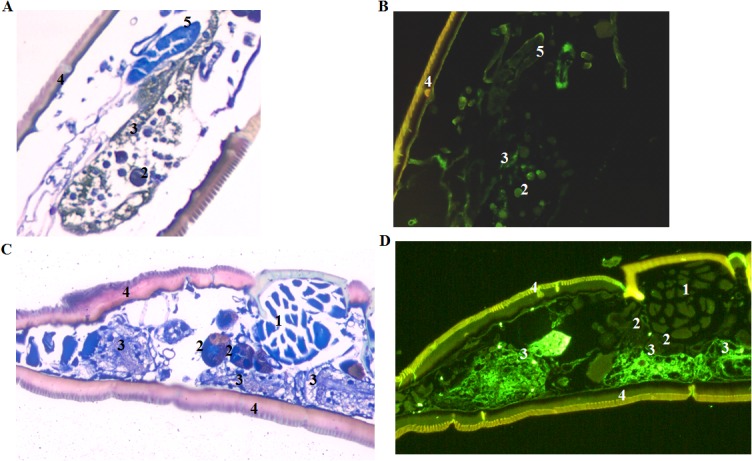
Fig 3. Visualization of LVS in colonized nymphs at 40 and 168 days post-capillary tube feeding.

(A) Wright Giemsa stained section of an LVS colonized nymph which was capillary tube fed LVS as a larva, molted to nymph, and was held at 22±1°C for 40 days. (B) Immunostained section of the same nymph. LVS colonization of both the salivary glands and gut was seen. (C) Wright Giemsa stained sections of an LVS colonized nymph which was capillary tube fed LVS as a larva, molted to a nymph, and was held at 22±1°C for 168 days. (D) Immunostained section of the same nymph. LVS colonization was observed only in the gut. Numbers identify (1) muscle, (2) salivary gland, (3) gut, (4) exoskeleton and (5) Malpighian tubule. 200x magnification.
