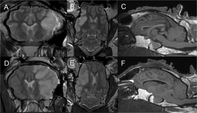
Fig 3. MRI findings at 24 hours following stroke: T1 and T2 weighted imaging.
Temporary MCAO demonstrates cerebral edema in a right MCA distribution similar to the diffusion deficit in Fig 2B on T2 coronal imaging (A), and no mass effect or midline shift on T2 axial imaging (B). Sagittal T1 sequences show preserved basal cisterns and posterior fossa CSF spaces (C). T2 coronal (D) and axial (E) imaging after permanent MCAO show cerebral edema distributed as for the diffusion deficit in Fig 2D, with associated mass effect and midline shift. Sagittal T1 imaging demonstrates effacement of the basal cisterns and cisterna magna, tonsillar herniation and brainstem compression (F). MCA, middle cerebral artery; MCAO, middle cerebral artery occlusion, n = 6/gp.

