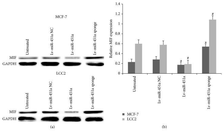
Figure 7.
Expression of MIF in each group. The MCF-7 and LCC2 cells were transfected with Lv-miR-451a or Lv-miR-451a sponge or Lv-miR-451a NC. Total lysates were prepared and subjected to western blot with anti-MIF antibody. # P < 0.05 versus Lv-miR-451a NC or control (MCF-7); ∗ P < 0.05 versus Lv-miR-451a NC or control (LCC2) (note: NC means negative control; Lv means lentivirus).

