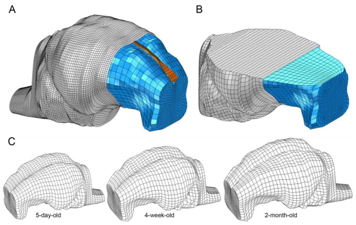
Figure 1.
(A) Whole brain FEM and (B) transection of the FEM for the 5-day-old piglet. Brain (blue), falx (orange), and skull (gray, A) or skull-PMMA- Plexiglas plate complex (gray, B). A portion of the frontal skull in each figure is cut away for illustration. (C) Isolated brain FEM for 5-day, 4-week, and 2-month-old piglets.

