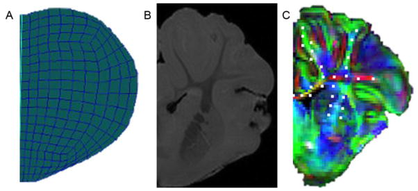
Figure 5.

(A) Right half coronal slice of FEM. (B) Right half coronal slice of high-resolution MRI scan. (C) Right half coronal slice of diffuse tensor image with overlaid RGB color map representing tissue tract orientation (red: x-direction, green: y-direction, blue: z-direction). Mesh node locations corresponding to WMTs are marked with white dots.
