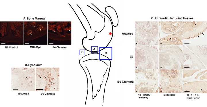
Fig 2. Immunostaining of MRL-specific markers in B6 chimeric mice.
Knee joint tissues of MRL control, B6 control and B6 chimeric mice are immunopositive for Class 1 MHC H2Kk (arrows indicate typical immunopositive cells). Location of the cartilage injury in the femoral groove is indicated by the red asterisk. Scale bars all = 100μm. A. Bone marrow: engraftment of MRL/MpJ bone marrow into B6 chimeric mice shown by the presence of MHC H2Kk-immunopositive cells (arrows). B: MRL-derived H2Kk-positive cells are seen in the synovium of B6 chimeric mice (arrows); C: MRL-derived cells are present in bone marrow of B6 chimeras but not in cartilage or other intra-articular tissues (bottom right panel).

