Figure 1.
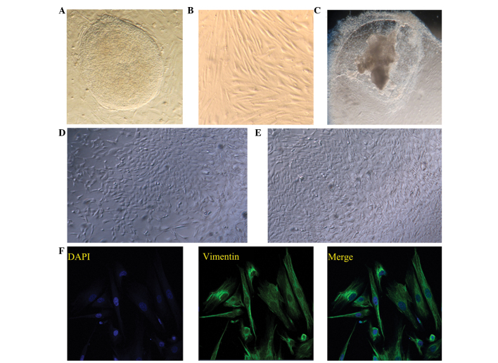
Induction of human embryonic stem cell (hESC) differentiation. Typical morphology of (A) hESC clone and (B) endometrial stromal cells. Images of the differentiated cells on day 21 following induction with (C) embryoid body, (D) mouse embryonic fibroblast feeder cells and (E) co-culture with endometrial stromal cells. Images (A–E) were captured using a Leica inverted microscope (magnification, x200). (F) Immunostaining of vimentin in the endometrial stromal cells, where 4′,6-diamidino-2-phenylindole was used to stain the nuclei. Fluorescent images were captured using an LSM 700 inverted confocal microscope (magnification, x400).

