FIGURE 5.
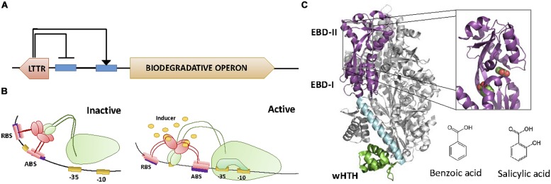
LysR family. (A) Architecture of the LTTR regulator and the regulated operon. LTTRs repress their own synthesis, while activating the transcription of the biodegradative operon. (B) Activation mechanism. Each of the dimers of the tetramer LTTRs binds to the RBS and the activating binding site (ABS), respectively. This causes the DNA to bend, preventing the access of the α-CTD of the RNApol to the promoter. Upon binding to the effector, the protein slides from ABS to ABS″. This movement directs the α-CTD of RNApol to contact DNA promoting transcription. (C) LTTR structure. The crystal structure of the LTTR CnbR (1IZ1, Muraoka et al., 2003) is shown. The inset shows the location of two benzoic acid molecules within BenM (2F78, Ezezika et al., 2007). The molecular structure of effectors benzoic acid and salicylic acid is also shown.

