FIGURE 7.
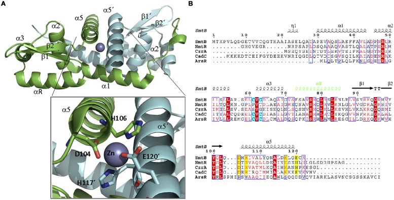
ArsR family. (A) ArsRF structure. The crystal structure of the dimer SmtB bound to Zn (II) (1R23, Muraoka et al., 2003) is shown. SmtB monomers are shown in green and blue, respectively. The inset shows the residues within α5 involved in metal binding. (B) ArsR family alignment. The alignment of ArsR members of Table 2 is shown. Residues of α3 or α5 involved in metal binding are highlighted in blue and yellow, respectively. DNA binding αR is shown in green.

