FIGURE 9.
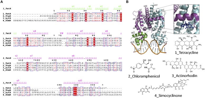
TetR family. (A) TetR family alignment. The alignment of representative TetR members for which effectors are antibiotics is shown. Secondary structure elements forming the DBD and the EDB are colored green and magenta, respectively. Alpha helices described in the text are labeled in the structure and in the alignment. (B) TFR structure. The crystal structure of the TFR TetR bound as a dimer to DNA (1QPI, Orth et al., 2000) is shown. The inset shows the location of a tetracycline derivative (iso-7-chlortetracycline) bound to TetR EDB (2X9D, Volkers et al., 2011). The molecular structure of antibiotic effectors recognized by the aligned proteins is also shown.

