
Fig. 10.
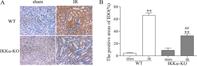
IKKα regulates the expression of IDO in kidneys after renal IR injury. C57BL/6 (wild type) or IKKα-null mice (IKKα-KO, IKKαfl/fl, Cre+/− mice) underwent sham-treated operation or ischemia-reperfusion by unilateral renal pedicle clamping for 45 min, followed by reperfusion (IR). The kidney tissues were harvested on day 3. (A) Representative images of IDO infiltrated the interstitium and renal TECs of IR kidneys. Kidneys of wild-type mice had intense and diffuse IDO staining within the tubule interstitium. However, enhancement of IDO expression is mediated by IKKα, as evidenced by minimum expression of IDO in IKKα-KO mice. (B) The IDO-positive areas were determined in a blind manner. Data are presented as mean±s.d. (n=6 mice). **P<0.01 versus respective sham treated; ##P<0.01 versus WT-IR.