
Fig. 3.
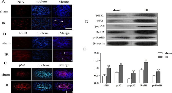
The expressions of NFκB cascade components in the repair phase of renal IR injury. C57BL/6 mice underwent unilateral renal pedicle clamping for 45 min, followed by reperfusion. (A-C) The kidneys were harvested on day 3. Immunofluorescence staining for NIK (A), p52 (B) and RelB (C) was performed. Positive staining for NIK and RelB was distributed in the renal TECs and tubulointerstitium; the positive staining for p52 was located in the nucleus. The sham-treated kidney exhibited faint staining for these cytokines, whereas IR caused the stronger staining on day 3. Scale bars: 20 μm. Arrows indicate positive results. (D) Protein levels of NIK, p52, p-p52, RelB and p-RelB were measured by western blotting. (E) Expression levels of NIK, p52, p-p52, RelB and p-RelB in the kidney, after normalization to β-actin were all significantly decreased in the IR kidneys compared with sham-treated kidneys. Data are presented as mean±s.d. (n=6 mice). **P<0.01 versus the sham treated.