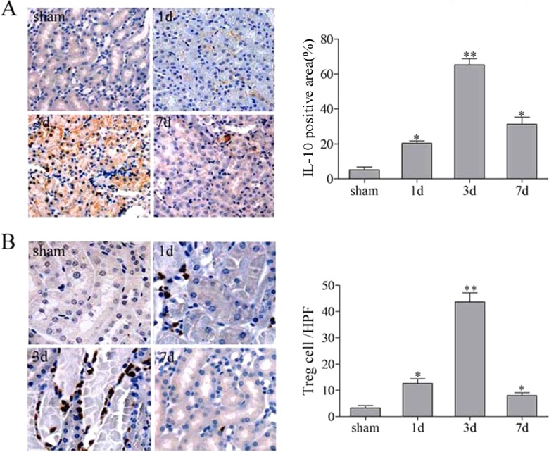
Fig. 6.

The expression of IL10 and Foxp3+ in the repair phase of renal IR injury. C57BL/6 mice underwent a sham-treated operation or unilateral renal pedicle clamping for 45 min, followed by reperfusion. The kidney tissues were harvested on days 1, 3 and 7. Representative immunehistochemistry staining of IL10 and Foxp3+ in the kidney from sham-treated or IR groups. (A) IL10 was located predominantly inside kidney vascular structures and in the interstitium, and was markedly increased on day 3. (B) The staining for Foxp3 was located in the nucleus with a peak on day 3. Data are presented as mean±s.d. (n=6 mice). *P<0.05 versus sham treated; **P<0.01 versus sham treated.
