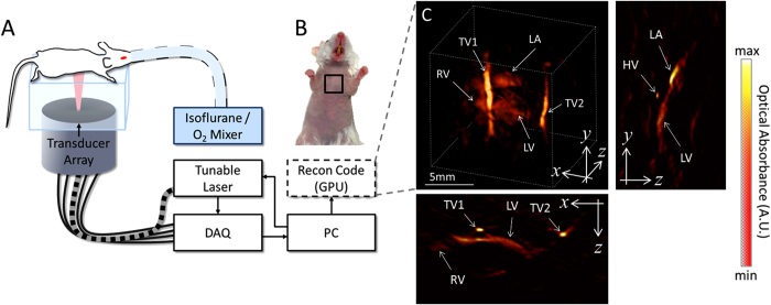
Figure 1. Experimental layout.
The experimental setup of the 4D optoacoustic tomography system is shown in panel A. The mouse was oriented in a prone position over the active surface of the ultrasound detector array. The heart of the mouse was illumated by shining laser light upon the chest via a fiber bundle connecting the tunable laser output to the center of the detector array. The imaged region corresponding to the live mouse heart is illustrated by the black square in panel B. A volumetric reconstruction of the heart and surrounding vessels is showcased in panel C. The cross-sections corresponding to the central position of the represented three-dimensional volume are also displayed in panel C. Important features of the heart, including the right ventricle (RV), left ventricle (LV), and left atrium (LA) are prominently visualized using the optoacoustic imaging approach. Additionally, two thoracic vessels (TV1 and TV2) can be clearly seen in the vicinity of the heart as well as a heart vessel (HV) indicated in a cross-section. The mouse and setup illustrations in A were developed by the authors.

