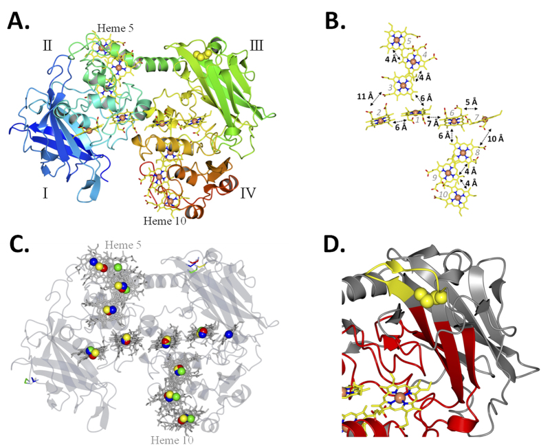
Figure 3. Crystal structure of MtrC at 1.8 Å resolution (PDB ID 4LM8).
(A) Cartoon representation of MtrC. The four domains indicated by roman numerals. The polypeptide chain is shown in cartoon representation and coloured from blue (N-terminus) to red (C-terminus). The iron atoms of the hemes are represented as orange spheres and the porphyrin rings of the hemes are shown as yellow sticks. The cysteines of the disulfide bond are represented as yellow spheres. (B) Heme packing and putative electron transfer distances between porphyrin rings (C) Alignment of iron atoms from MtrC (yellow) over the iron atoms of MtrF (red), OmcA (green) and UndA (blue). The transparent structure of MtrC is shown in grey as (D) Domain III of MtrC showing the position of the disulfide (yellow spheres). Residues within 16 Å of heme 7 are shown in red.

