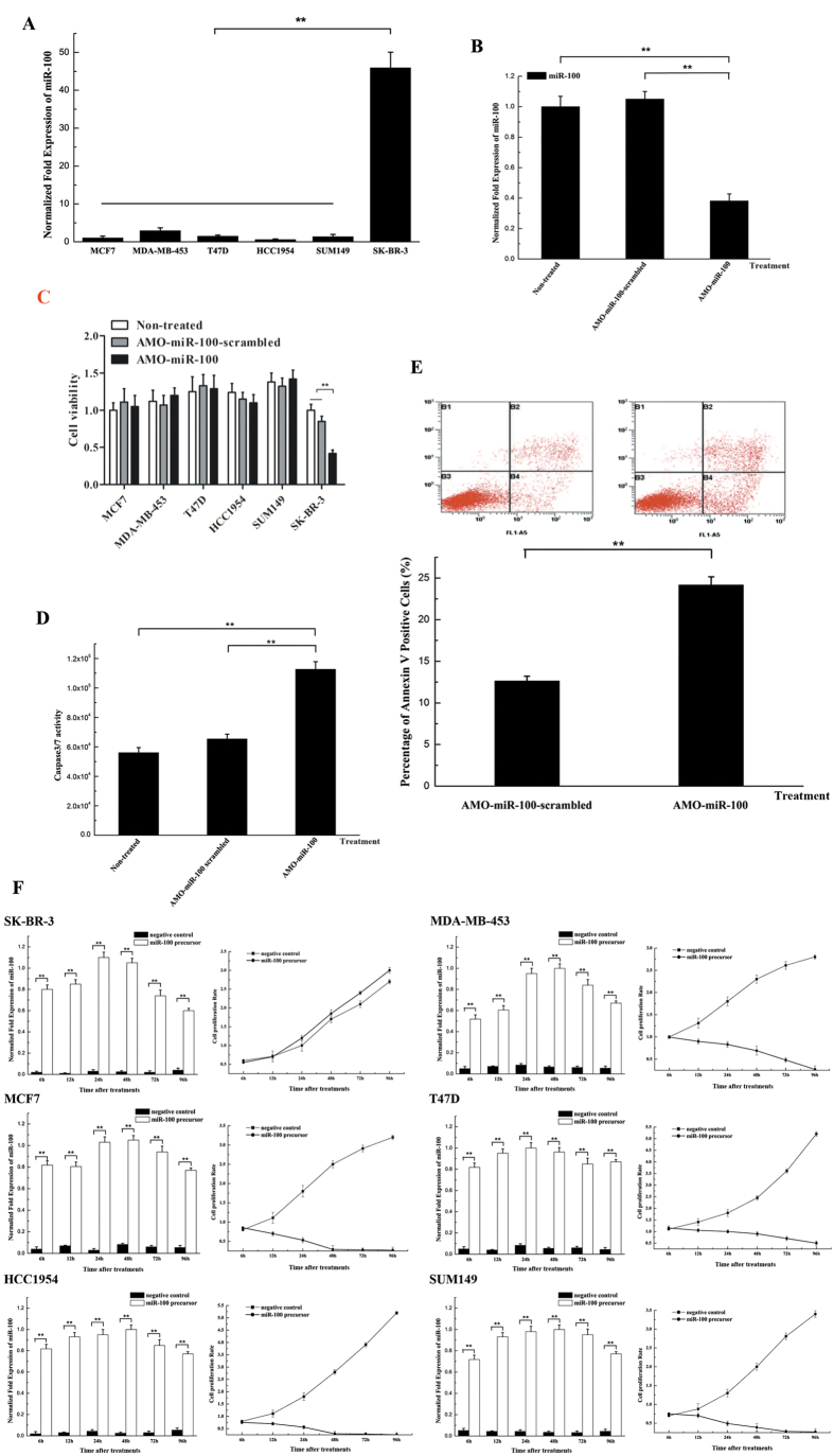
Figure 1. The role of miR-100 in the regulation of apoptosis in breast cancer cells.
(A) The expression of miR-100 in breast cancer cells. The expression level of miR-100 in different cell lines including SK-BR-3, T47D, SUM149, HCC1954, MDA-MB-453 and MCF7 was examined with quantitative real-time PCR. (B) Silencing of miR-100 expression in breast cancer cells. AMO-miR-100 or AMO-miR-100-scrambled was transfected into SK-BR-3 cells, followed by detection of miR-100 expression using real-time PCR at 48 h after transfection. Non-treated cells were used as controls. (C) The effect of miR-100 silencing on cell viability. Cell viability was evaluated at 48 h after transfection of breast cancer cells with the AMOs. (D) Influence of miR-100 silencing on caspase 3/7 activity in breast cancer cells. The SK-BR-3 cells at 36 h after transfection of AMO-miR-100 or AMO-miR-100-scrambled were subjected to the Caspase-Glo 3/7 assay to evaluate apoptosis. (E) The effects of miR-100 downregulation on apoptosis using Annexin V assays. Apoptosis was examined by flow cytometry at 48 h after transfection of AMOs. (F) Overexpression of miR-100 in breast cancer cells. SK-BR-3, MCF7, HCC1954, MDA-MB-453, T47D and SUM149 cells were transfected with the miR-100 precursor or a negative control. At different times after transfection, the cells were subjected to real-time PCR to detect miR-100 and the effects of miR-100 overexpression on cell proliferation were analysed. In all panels, plotted data referred to the means ± standard deviations of triplicate assays and asterisks represented statistically significant differences (**p < 0.01).

