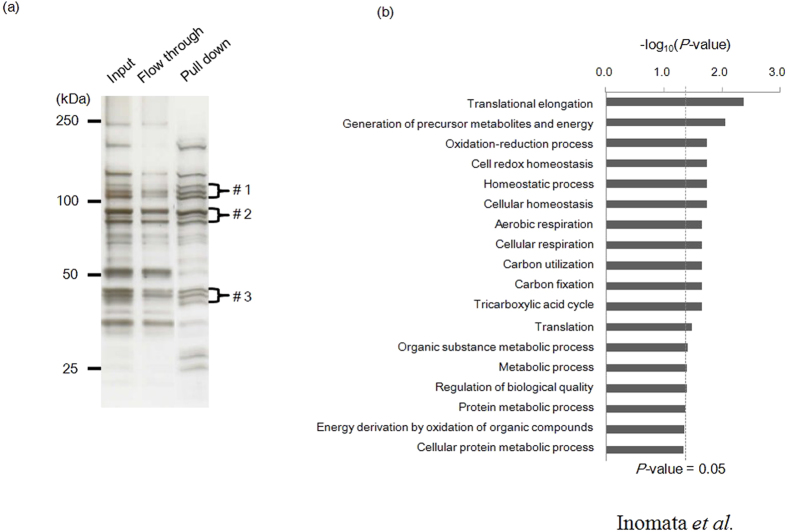
Figure 4. Identification of heparin-binding proteins of C. parvum sporozoites.
(a) Silver staining showing the whole lysates of C. parvum sporozoite (lane 1, Input), heparin-unbound proteins (lane 2, Flow through), and heparin-binding proteins (lane 3, Pull down). The proteins in the bands with molecular masses of 120 (#1), 90 (#2), and 45 (#3) kDa were specifically concentrated in the precipitated fractions, and proteins in these three bands were separately gel-extracted and subjected to mass spectrometry analysis. (b) Gene enrichment analysis of heparin-binding proteins. To functionally categorize the proteins that interacted with heparin, all of the proteins identified by mass spectrometry were assigned to a GO grouping. GO analysis was carried out by using Gene Ontology Enrichment embedded in the CryptoDB database (http://cryptodb.org/), where Fisher’s exact P values were used to determine the GO terms that were statistically significant (P < 0.05).

