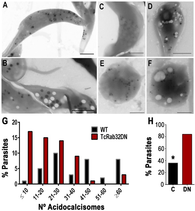
Fig. 7.

Reduction in electron-dense acidocalcisomes and a considerable increase in empty vacuoles in cells expressing dominant-negative TcRab32 in comparison with wild-type cells. (A,B) Direct transmission electron microscopy image from whole unstained and unfixed epimastigotes expressing dominant-negative GFP–TcRab32 (DN) show the presence of numerous empty vacuoles (B) in comparison with wild-type epimastigotes (A). (C–F) A similar increase in the number of empty vacuoles was observed in trypomastigotes (C,D) and amastigotes (E,F) expressing dominant-negative GFP–TcRab32 (D,F) as compared with that of wild-type cells (C,E). (G) The number of acidocalcisomes per epimastigote was counted in 70 random cells from two independent experiments, and the numeric distribution of acidocalcisomes showed that the majority of epimastigotes expressing dominant-negative TcRab32 (TcRab32DN) had <10 or between 11 and 20 electron-dense acidocalcisomes. The results in wild-type (WT) cells are similar to those reported previously for trypanosomatids. (H) An increase in the percentage of epimastigotes showing empty vacuoles. In order to quantify the phenotype of empty vacuoles, by using transmission electron microscopy, we examined 50 random wild-type parasites, ‘C’, and parasites expressing the dominant-negative protein (DN), and counted the number of parasites with empty vacuoles for each. In comparison with wild type, we found that there was a significant increase in the percentage of parasites with empty vacuoles when they expressed dominant-negative TcRab32. *Differences between control and the dominant-negative protein are statistically significant, P<0.05. Scale bars: 2 µm (A,B); 1 µm (C–F).
