Figure 1.
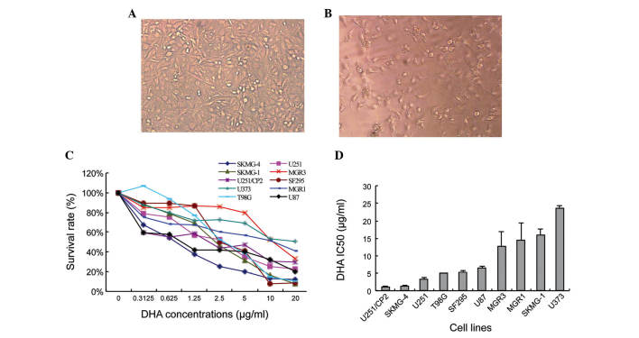
DHA exhibits cytotoxicity in glioma cell lines in vitro. The density and cellular morphology of SKMG-4 cells cultured (A) without and (B) with DHA (magnification, ×200). The cells appeared lower in density and shrunken following treatment with DHA. (C) Survival curve of the 10 human glioma cell lines following treatment with serial concentrations of DHA for 72 h. (D) IC50 of the 10 glioma cell lines. Values are expressed as the mean ± standard deviation. DHA, dihydroartemisinin; IC50, half maximal inhibitory concentration.

