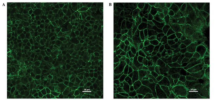
Figure 3.
Subcellular localization of claudin-3 in breast cancer cell lines visualized by immunofluorescence. Breast cancer cells lines (A) MCF-7 and (B) MDA-MB-415 (overexpressing claudin-3) were plated onto chambered cover slides and incubated with an antibody to claudin-3. Claudin-3 protein was primarily observed at cell junctions but was also detected intracellularly (to a lesser extent) in both cell lines.

