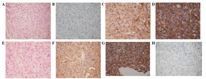
Figure 3.
Immunohistochemical staining in the resected tumors of case one and two. Case one. (A) The tumor consisted of highly spindle cells in a dense hyalinized collagenous stroma, which consisted of alternating hypercellular and hypocellular areas, while thin-walled staghorn branching vessels were identified (hematoxylin and eosin staining; magnification, ×200). (B) Ki-67 demonstrated scattered positivity (2% of tumor cells) (magnification, ×200). Immunohistochemically, the tumor cells exhibited strong positive staining for (C) vimentin (magnification, ×400) and (D) CD34 (magnification, ×400). Case two. (E) The tumor was composed of spindle cells interspersed between dense collagen bundles and vascular channels, without atypia (hematoxylin and eosin staining; magnification, ×200). Immunohistochemically, the tumor cells exhibited positive staining for (F) CD34 (magnification, ×400) and (G) vimentin (magnification, ×400) and negative staining for (H) epithelial membrane antigen (magnification, ×200). CD, cluster of differentiation.

