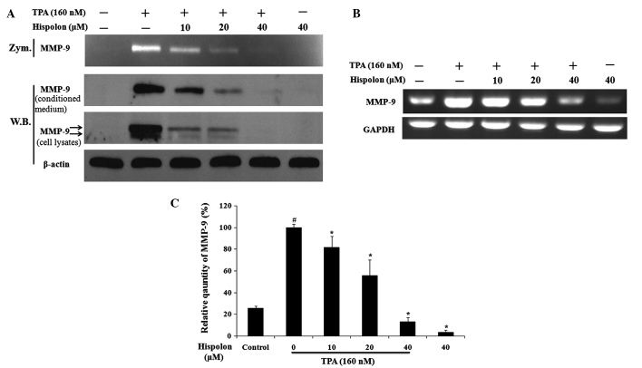
Figure 3.
Hispolon inhibited TPA-induced MMP-9 activation, as well as inhibited the secretion and expression in a dose-dependent manner. (A) Cells in serum-free medium were pretreated with hispolon at the indicated concentrations for 2 h prior to TPA incubation for another 24 h. Conditioned media were analyzed by gelatin zymography (upper panel) and western blot analysis (middle panel). Cell lysates were collected and subjected to western blot analysis (lower panel). β-actin was used as an internal control. (B) Hispolon inhibited TPA-induced MMP-9 gene expression. MMP-9 expression was detected by RT-PCR and GAPDH was used as an internal control. (C) Relative quantification of MMP-9 gene expression. All the data are presented as the mean ± standard deviation of three independent experiments. *P<0.05 vs. TPA alone-treated group; #P<0.05 vs. DMSO control. TPA, 12-O-tetradecanoylphorbol-13-acetate; MMP-9, matrix metalloproteinase-9; RT-PCR, reverse transcription-polymerase chain reaction; DMSO, dimethyl sulfoxide.

