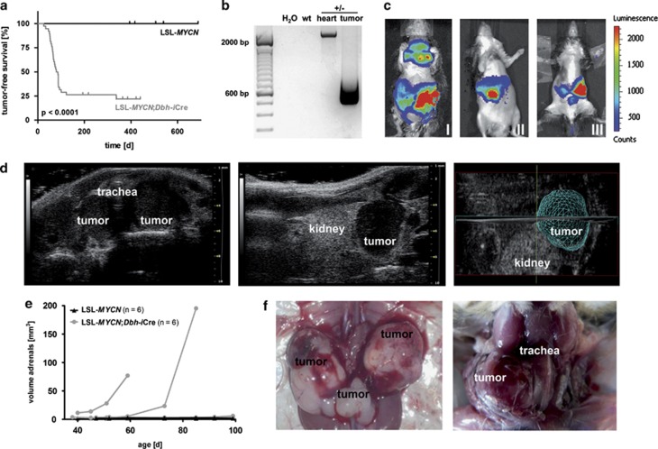
Figure 2.
Double-transgenic LSL-MYCN;Dbh-iCre mice develop tumors derived from the neural crest. (a) Kaplan–Meier analysis indicating the presence and the time to detection of palpable tumors in mice (that is, tumor-free survival) heterozygous for LSL-MYCN and mice double transgenic for LSL-MYCN and Dbh-iCre (Log-rank test). (b) Representative result of PCR validating the removal or presence of the transcriptional termination site 5′ to the MYCN transgene in tumor and control tissues, respectively. Wild type (wt), double-transgenic LSL-MYCN;Dbh-iCre (+/−). (c) Bioluminescence imaging of three representative LSL-MYCN;Dbh-iCre mice carrying palpable tumors at the superior cervical ganglion (I), adrenals (I, II, III) or celiac ganglion (III). Color code indicates luciferase activity (low=blue; high=red). (d) High frequency ultrasound images of palpable tumors arising from superior cervical ganglion (left) and adrenal (middle), and three-dimensional reconstruction of adrenal tumor (right). (e) Growth curves of tumors, as detected by high-frequency ultrasound. (f) Macroscopic images during autopsy of mice carrying palpable tumors arising from both adrenals and the celiac ganglion (left) and from the superior cervical ganglion (right).

