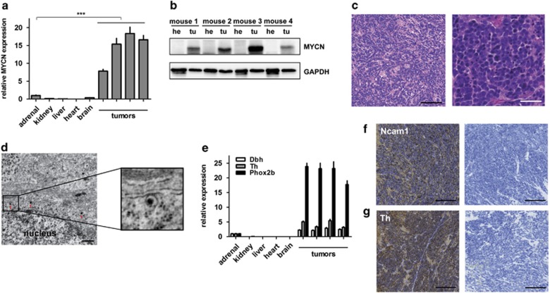
Figure 3.
Tumors of LSL-MYCN;Dbh-iCre mice resemble human neuroblastoma in terms of histology and molecular expression patterns. (a) MYCN expression (qPCR) in four representative tumors from LSL-MYCN;Dbh-iCre mice compared with control tissues. Expression was normalized to normal adrenal glands. Student's t-test: ***P<0.001. (b) Western blot analysis confirms MYCN expression in tumors (tu) compared with heart (he) tissue collected from four representative double-transgenic mice. (c) Hematoxylin and eosin (H&E) staining shows small, round blue cells typical for neuroectodermal tumors. Scale bars=100 μm (left) and 50 μm (right). (d) Electron micrographs show neuronal structures, including neurosecretory vesicles (red arrows). Scale bar=500 nm. (e) Reverse transcription-qPCR confirms significantly increased expression of the murine orthologs of the human neuroblastoma marker genes dopamine β-hydroxylase (Dbh), tyrosine hydroxylase (Th) and paired-like homeobox 2b (Phox2b) in tumors compared with normal control tissues (Student's t-test; Dbh: P=0.005, Th: P=0.02, Phox2b: P=0.0006). (f, g) Immunohistochemistry confirms expression of neuroblastoma markers, Ncam1 and Th. Scale bars=200 μm.

