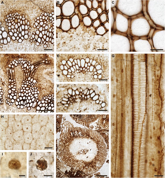
Fig. 4.

Anatomical and cytological details of Osmunda pulchella sp. nov. from the Lower Jurassic of Skåne, southern Sweden (A–F, I: NRM-S069656; G, H, J, K: NRM-S069658). a Detail showing pith parenchyma (bottom), stelar xylem cylinder dissected by complete leaf gaps, triangular section of phloem projecting into leaf gap, and parenchymatous inner cortex (top); note mesarch leaf-trace protoxylem initiation in the stelar xylem segment on the right. b Detail of (a) showing peripheral pith parenchyma and stem xylem. c Detail of stem xylem showing tracheid pitting. d Endarch leaf trace emerging from the stele and associated with a single root. e Leaf trace in the inner cortex of the stem showing single, endarch protoxylem cluster. f Leaf trace immediately distal to initial protoxylem bifurcation in the outermost cortex of the stem. g Root vascular bundle showing well-preserved scalariform pitting of metaxylem tracheids. h Well-preserved pith parenchyma showing membrane-bound cytoplasm with cytosol particles and interphase nuclei containing nucleoli. i, j Nuclei with conspicuous nucleoli (in interphase: I) or with distinct chromatid strands (in prophase: J). k Transverse section through root showing diarch vascular bundle, parenchymatous inner cortex with isolated fibre strands, and prominent fibrous outer cortex. Scale bars: (a) = 100 μm; (b, e, f, h) = 50 μm; (c, g) = 25 μm; (d) = 200 μm; (i, j) = 5 μm; (k) = 250 μm
