Fig. 5.

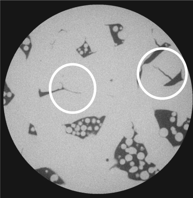
Sample 1–5D, X-ray CT of the interior of a 5 mm diameter cylinder cut from the original sample. The 5 mm cylinder had the same height as the original sample, but the image stack only extends part way through the sample. The pixel size in this image is 0.87 µm. Table 7 gives more details about the images taken from this sample. The white circles mark apparent cracks.
