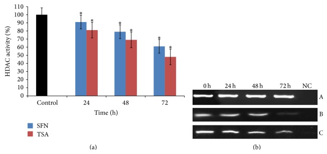
Figure 2.

Effect of SFN and TSA on HDAC1 in human cervical cancer cells (HeLa). (a) 2.5 μM of SFN and 0.05 μM TSA treatments significantly inhibit the activity of HDAC in time-dependent manner, respectively. Values are means ± SD of three independent experiments. Symbol (∗) indicates significant (P < 0.05) difference of data between control and treated cells. (b) 2.5 μM SFN treated HeLa cells show significantly time-dependent reduction in the mRNA expression of HDAC1 in comparison to untreated cells. Panel A shows β-actin expression as an internal control, Panel B shows the expression of HDAC1 on treatment with TSA, and Panel C shows the expression of HDAC1 on treatment with SFN. Lane 1 shows the expression of HDAC1 gene in untreated HeLa cells; Lanes 2, 3, and 4 show the time-dependent decrease in the expression of HDAC1 after treatment for 24, 48, and 72 h, respectively; Lane 5 shows negative control for RT-PCR.
