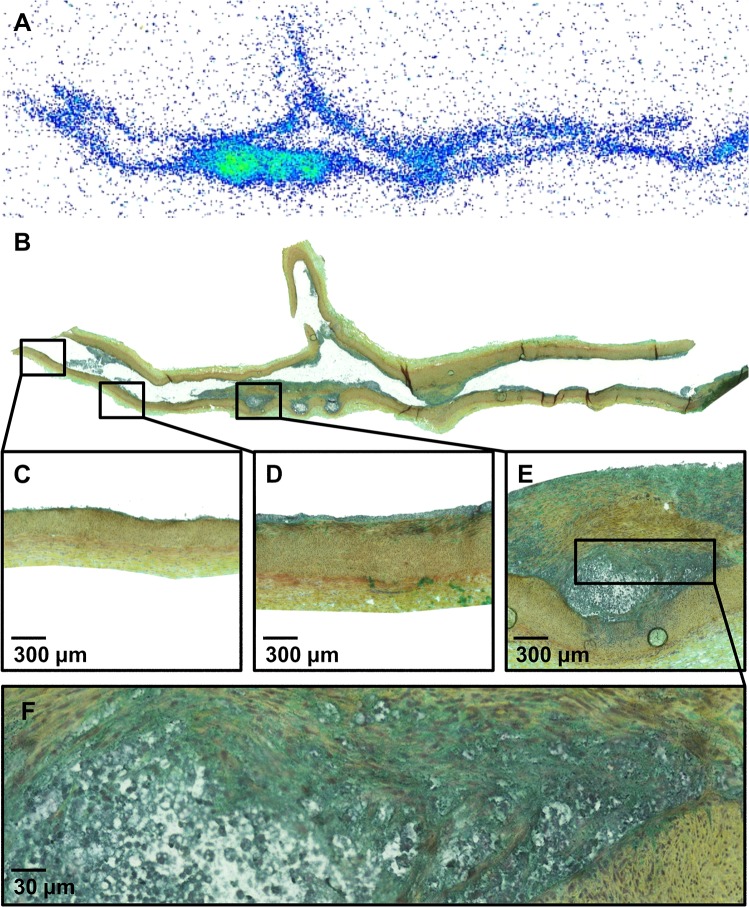
Fig 2. Representative [18F]FDG autoradiograph (A) and a serial tissue section stained with Movat pentachrome (B).
The inserts show areas of normal vessel wall (C), intimal thickening (D) and atheroma (E) at higher magnification. Panel F shows lipid core surrounded by densely cellular tissue at high magnification.

