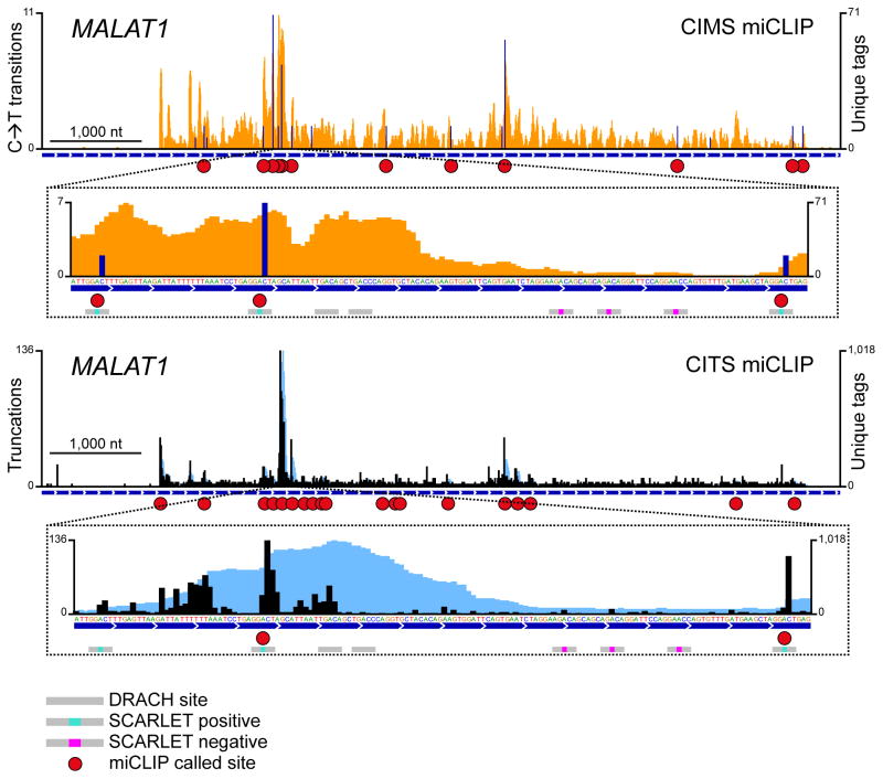
Figure 3. miCLIP identifies m6A with single-nucleotide resolution.
m6A residues detected by CIMS and CITS miCLIP in the ncRNA MALAT1. Orange and dark blue tracks: CIMS miCLIP unique read coverage and C→T transitions, respectively. Light blue and black tracks: CITS miCLIP unique read coverage and unique read starts, respectively. Horizontal blue bars: Transcript models. Red circles: miCLIP called m6A. Small horizontal bars in insets: DRACH consensus sites with a methylation status that is undefined (grey), confirmed positive (turquoise) or confirmed negative (magenta) by SCARLET19.

