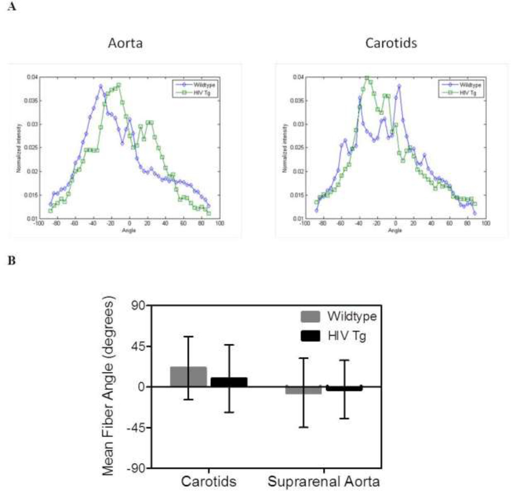
Figure 5. Collagen fiber orientation is not different between groups.
The distribution of collagen fibers within the adventitia was determined using fast Fourier transform techniques on images from the two-photon microscopy z-stacks. Panel A shows the average distributions across the wall for the carotids and aortas. Panel B plots the weighted mean fiber angles which show no statistically significant differences are between the HIV Tg and wildtype mice (N=4, p<0.05).

