Fig. 2.

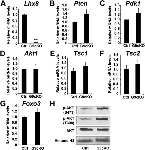
AKT is activated in Lhx8 flx/flx Gdf9Cre oocytes. a–g Oocytes were isolated from PD7 control (Lhx8 flx/flx, Ctrl) and Lhx8 flx/flx Gdf9Cre (G9cKO) mouse ovaries and RNA was extracted for cDNA conversion and real-time quantitative polymerase chain reaction (RT-qPCR). Data were normalized to Gapdh expression and are given as the mean relative quantity (compared with control), with error bars representing the standard error of the mean. Student’s t-test was used to calculate P values. The only significant difference was noted in the expression of Lhx8, as expected. ** P < 0.01. h Oocytes were isolated from PD7 control and Lhx8 flx/flx Gdf9Cre ovaries, protein was extracted, and a Western blot test was performed on three independent samples, using antibodies against AKT and its two phosphorylated forms (S473 and T308). Histone H3 immunoreactivity was used as a control
