Fig. 6.

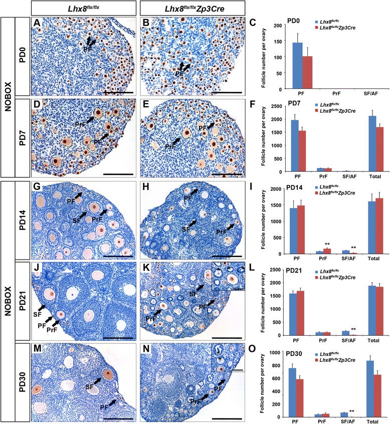
Lhx8 inactivation in primary follicles (Lhx8 flx/flx Zp3Cre) abolishes follicle growth. Histomorphological analysis was done on control (Lhx8 flx/flx) and Lhx8 deficient ovaries (Lhx8 flx/flx Zp3Cre) at various stages of postnatal ovarian development ranging from newborn (PD0) to postnatal day 30 (PD30). a–f Periodic acid–Schiff (PAS) staining and counting of different follicle types in the newborn and PD7 ovaries showed no significant differences between control and Lhx8 deficient ovaries. g–o Anti-NOBOX antibodies were used to stain oocytes (brown immunoreactivity) in ovaries from PD14 (G and H), PD21 (J and K), and PD30 (M and N) mice. At PD14, the Lhx8 flx/flx Zp3Cre ovaries showed a significantly higher number of primary follicles (PrF) and significantly diminished number of secondary/preantral (SF/AF) follicles characterized by two or more layers of granulosa cells. At PD21 and PD30, the primary follicle pool did not differ significantly between Lhx8 flx/flx Zp3Cre and control ovaries; however, there was a marked decrease in the number of secondary and more advanced ovarian follicles in conditional knockouts including degenerating follicles without oocytes (marked by asterisks in insets in K and N). The primordial follicle (PF) pool remained relatively stable between PD14 and PD30, with no significant difference between Lhx8 flx/flx Zp3Cre and control ovaries. **P < 0.01. Scale bars: 100 μm (A, B, D, and E); 200 μm (G, H, J, K, M, and N); 50 μm (insets of K and N)
